Your orthodontic workup: what it is and why it matters
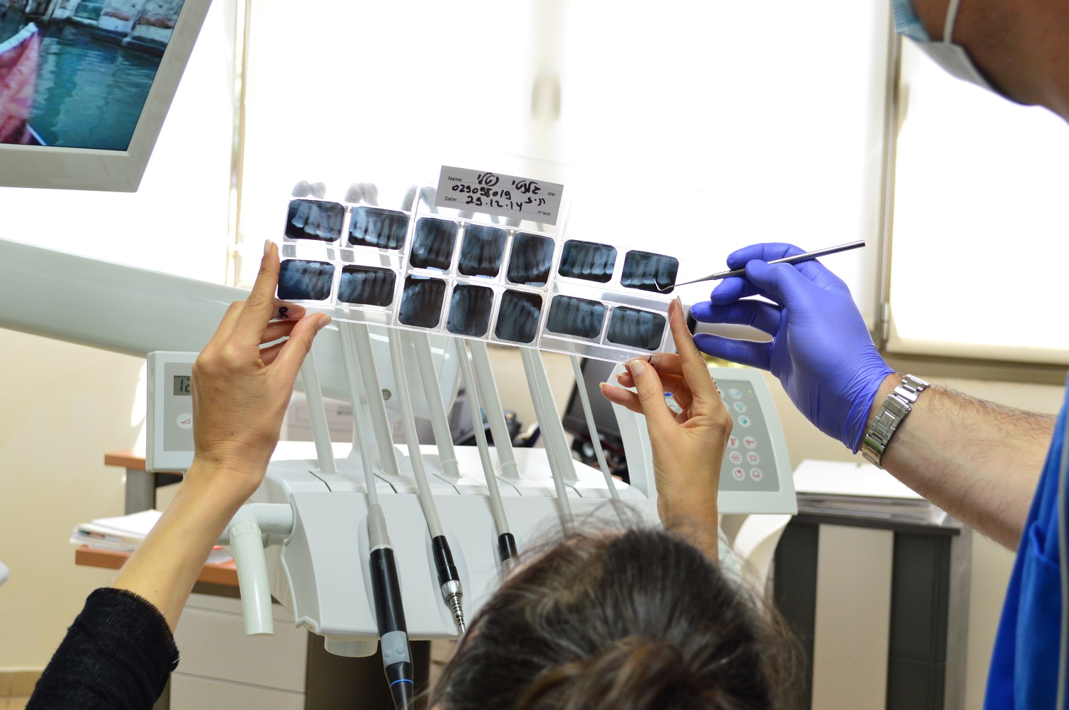
Before we put on any appliance — whether aligners or braces — we do a thorough study of your mouth, teeth, and facial bones. For many patients, this step raises questions: why do we need so many photos and x-rays? Is it really necessary?
Yes, it is. And this guide explains exactly what’s involved and why it makes all the difference.
What’s the point of the orthodontic study?
Think of it this way: no one starts building a house without blueprints. Your orthodontic workup is our set of blueprints.
It lets us see what can’t be seen with the naked eye: how the bones are positioned, how much space the teeth have to move, whether there are teeth that haven’t erupted yet, how deep the bite is, and whether there are growth factors we can take advantage of.
With that information we design a treatment plan tailored specifically to you — not a generic one. And we can anticipate situations that would otherwise be unpleasant surprises mid-treatment.
Patients who go through a complete workup tend to have:
- More efficient treatments that finish faster
- Fewer emergency or unplanned appointments
- More stable, longer-lasting results
- Greater predictability in timing and cost
What does the study include?
Clinical photographs
We take two types of photos:
Intraoral photos (inside the mouth): from the front, from the right and left sides, and from above and below each arch. They show crowding, the bite, overall tooth condition.
Extraoral photos (of the face): at rest from the front, smiling from the front, from each profile, and in three-quarter view. We assess facial symmetry, lip position, and facial profile.
These photos also serve as a record of your progress throughout treatment — comparing before and after is always motivating.
Study models
These are exact reproductions of your teeth and gums. They can be made two ways:
- Traditional method: impressions with a dental material, then plaster replicas
- 3D intraoral scanner: we pass a small scanner over your teeth, and in a few minutes we have a perfect digital model — no uncomfortable impression material needed
The models let us analyze the shape of your arches, measure available space, study the bite from every angle, and plan exactly which movements are needed.
Panoramic x-ray
A single image that shows your entire mouth: all your teeth (including those that haven’t erupted), the jaw bones, the jaw joint, and the sinuses. In one image we can see impacted teeth, overall bone health, or anything that needs attention before treatment begins.
Cephalometric x-ray (lateral skull film)
This is a side-view x-ray of the full head. From it we make precise measurements — cephalometry — that tell us:
- How the upper jaw and lower jaw relate to each other
- The angle at which the teeth are positioned relative to the bones
- Whether there’s a skeletal discrepancy (one bone more forward or back than the other)
- Lip position (whether they protrude or sit back)
- Any asymmetries to consider
These measurements are compared to established norms for your age and background, and they guide our planning.
Cone beam CT scan (CBCT)
Not every patient needs this, but when it’s indicated, it’s an extraordinary tool. It produces a full 3D image of your facial structures.
We use it for special cases: teeth impacted in complex positions, mini-implant planning, airway analysis (important for sleep apnea risk), or preparation for orthognathic surgery.
Does any of it hurt?
Nothing in the study is painful. Photos are photos. X-rays are quick and you don’t feel them. The intraoral scanner is like having a small wand passed over your teeth.
If we do traditional impressions, some patients feel a mild discomfort from the material, but it’s brief.
What happens after the study?
Once we have all the records, we analyze them carefully and prepare your personalized treatment plan. At your next appointment we walk you through everything: what we found, what can be corrected, how long we estimate treatment will take, and we answer every question you have.
Nothing begins until you’re fully informed and on board.
The orthodontic workup isn’t a bureaucratic formality — it’s the map that gets us to your ideal smile as safely and efficiently as possible.
Ready to take the first step? Book your diagnostic consultation and let’s get to know your case.